August 30, 2012
What is the Evidence for Induction for Low Amniotic Fluid in a Healthy Pregnancy?
By: Sharon Muza, BS, LCCE, FACCE, CD/BDT(DONA), CLE | 0 Comments
By Rebecca L. Dekker, PhD, RN, APRN
Today's post on the Evidence for Induction for Low Amniotic Fluid in a Healthy Pregnancy is a guest post by blogger Rebecca Dekker, owner of the fairly new blog in the birth world, Evidence Based Birth that has been very well received and enjoyed by many. Look for an interview with Rebecca in an upcoming post where we will learn how this Assistant Professor of Nursing who teaches pathopharmacology and studies depression in patients with heart failure ended up writing the Evidence Based Birth blog appreciated by birth professionals. I look forward to future posts and collaboration with Rebecca and thank her for her contribution today.- SM
__________________
This question came from one of my readers:
'Low fluid seems to be the new 'big baby' for pushing for induction. What does the research say about low fluid at or near term? From what I've been able to see in research summaries at least, there appears to be no improved outcome for babies, but I'd love to see the research really hashed out. I'm also curious about causes of low fluid (theorized or known), risks of low fluid, and perhaps as important if not more so, measurements of low fluid.'
This is a great question and I felt like it was a perfect topic for my first article for Science and Sensibility. Standard of practice in the U.S. is to induce labor at term if a mother has low amniotic fluid in an otherwise healthy pregnancy. In fact, 95% of physicians who practice maternal-fetal medicine feel that isolated oligohydramnios - low amniotic fluid in an otherwise healthy pregnancy - is an indication for labor induction at 40 weeks (Schwartz, Sweeting et al. 2009).
But what is the evidence for this standard birth practice? Let's take a look at the evidence together.
First of all, what is oligohydramnios?
Oligohydramnios means low fluid inside the amniotic sac.
(oligo = little, hydr = water, amnios = membrane around the fetus, or amniotic sac).
Not sure how to pronounce oligohydramnios? Click here.

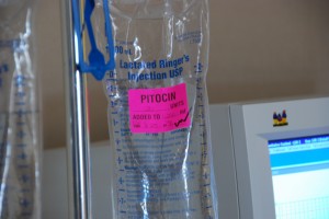
It is standard of care in the U.S. to induce women
with isolated oligohydramnios at term.
Image Source drewesque
What is amniotic fluid, and what does it do?
During pregnancy, the baby is surrounded by a liquid called amniotic fluid. Amniotic fluid helps protect the baby from trauma to the mother's abdomen. Amniotic fluid cushions the umbilical cord, protects the baby from infection, and provides fluid, space, nutrients, and hormones to help the baby grow (Brace 1997).
During the second half of pregnancy, amniotic fluid is made up of the baby's urine and lung secretions. This liquid originally came from the mother, and then flowed through the placenta, to the baby, and out through the baby's bladder and lungs (Brace 1997).
This same amniotic fluid is then swallowed by the baby and re-absorbed by the lining of the placenta. Because the mother's fluid levels are the original source of amniotic fluid, changes in the mother's fluid status can result in changes in the amount of amniotic fluid. Amniotic fluid levels increase until the mother reaches about 34-36 weeks, and then levels gradually decline until birth (Brace 1997).
What can cause low amniotic fluid at term?
Both mother and baby factors can contribute to low amniotic fluid at term.
Mother factors:
- If the mother is dehydrated, this may lower the amniotic fluid levels. (Patrelli, Gizzo et al. 2012)
- Women are more likely to be diagnosed with low amniotic fluid levels during the summer, possibly because of dehydration. (Feldman, Friger et al. 2009)
- If a woman with low amniotic fluid levels at term drinks at least 2.5 Liters of fluid per day, she increases the likelihood that her amniotic fluid levels will be back up to normal by the time of delivery. (Patrelli, Gizzo et al. 2012)
- If the mother rests on her left side before or during the fluid measurement, this can increase amniotic fluid levels. (Ulker, Temur et al. 2012)
- If the mother's water has broken (membranes ruptured), this will lead to a decrease in amniotic fluid. (Brace 1997)
- If the mother's placenta is not acting sufficiently anymore, this may lead to a decrease in amniotic fluid. When this happens, it may be because the mother has a serious condition such as pre-eclampsia or intrauterine growth restriction. (Beloosesky and Ross 2012)
Baby factors:
- If the baby has a problem with the urinary tract or kidneys, this may decrease the flow of urine. (Brace 1997)
- In the 14 days before the start of spontaneous labor, the baby's urine output starts to decrease. (Stigter, Mulder et al. 2011)
- As the baby gets closer to term, the baby swallows more amniotic fluid, thus leading to a decline in fluid levels. (Brace 1997)
- If the baby is post-term (after 42 weeks), he or she begins to swallow significantly more fluid, contributing to a decline in amniotic fluid. (Brace 1997)
- If the baby has a birth defect, he or she may swallow significantly more fluid, leading to low amniotic fluid levels. (Beloosesky and Ross 2012)
What is the best way to measure amniotic fluid levels?
The gold-standard method is to inject the amniotic sac with dye and then take samples of the amniotic fluid to check the dilution. However, this method is very invasive. So the most commonly used methods instead are 2 ultrasound techniques: the amniotic fluid index (AFI) and thesingle deepest pocket (Gilbert 2012).
To calculate the AFI, the technician divides the uterus into 4 areas. The largest fluid pocket in each area is measured, and then these 4 numbers are added make up the AFI. An AFI value of 5 cm or less is considered oligohydramnios. With the single deepest pocket method, the technician looks for the largest pocket of amniotic fluid in the uterus. If the largest pocket is less than 2 cm by 1 cm, then that is considered a diagnosis of oligohydramnios (Nabhan and Abdelmoula 2009).
It is important to understand that amniotic fluid levels exist on a continuum and that there is no agreement among researchers about the cut-off value that predicts poor outcomes - the AFI level of 5 was arbitrarily chosen to define oligohydramnios (Nabhan and Abdelmoula 2009). Furthermore, a large body of research has shown that both AFI and single deepest pocket are poor predictors of true amniotic fluid volume. For example, the AFI catches only 10% of all cases of true oligohydramnios (10% sensitivity)(Gilbert 2012).
There are several factors that make it difficult to get an accurate ultrasound measurement. As fluid levels decrease, ultrasound results become less accurate. Inexperience on the part of the technician can reduce the accuracy of the test results, as well as the amount of pressure that the technician puts on the ultrasound probe. The position of the baby can also affect the accuracy of the results. (Nabhan and Abdelmoula 2009; Gilbert 2012).
So which is the best way to measure amniotic fluid?
In a Cochrane review, researchers combined the results from 5 randomized controlled trials with more than 3,200 women. In these studies, women were randomized to either the AFI method or the single deepest pocket method. Researchers found that when the AFI is used to measure amniotic fluid, women were 2.4 times more likely to be diagnosed with oligohydramnios, 1.9 times more likely to be induced, and 1.5 times more likely to have a Cesarean for fetal distress without any corresponding improvement in infant outcomes. The researchers concluded that the single deepest pocket measurement has fewer risks and should be the preferred way to measure amniotic fluid (Nabhan and Abdelmoula 2009).
What is the clinical significance of low amniotic fluid when a mother reaches 37 or more weeks?
In 2009, 91% of physicians believed that isolated oligohydramnios, or low amniotic fluid in an otherwise healthy pregnancy at term, was a risk factor for poor outcomes (Schwartz, Sweeting et al. 2009).

In the U.S., 91% of maternal-fetal physicians
believe that isolated oligohydramnios at term is a
risk factor for poor outcomes, and 95% will
recommend labor induction.
Image Source robenjoyce
However, this belief is not accurate. In early studies on amniotic fluid and outcomes, researchers included babies with congenital defects , women with pre-eclampsia or intrauterine growth restriction (IUGR), and women who were post-term (past 42 weeks) in their samples. These women and babies are more likely to have low amniotic fluid, and they are also much more likely to have poor outcomes. So although early researchers found that babies born to women with low amniotic fluid had higher perinatal mortality rates (Chamberlain, Manning et al. 1984), higher Cesarean rates for fetal distress, and lower Apgar scores (Chauhan, Sanderson et al. 1999), the poor outcomes were due to the complications - not the low amniotic fluid (Gilbert 2012).
So, if a woman has TRUE ISOLATED oligohydramnios at term, meaning low amniotic fluid in a healthy pregnancy with a healthy baby at term (between 37 and 42 weeks), what are the risks?
There is no evidence that isolated oligohydramnios at term is a risk factor for poor outcomes. However, induction for isolated oligohydramnios leads to higher Cesarean rates. In a systematic literature review, I found 5 studies from the last 10 years. I will discuss the 3 highest quality studies here. For results from all 5, you can see my findings summarized in this Google document table here.
- Locatelli et al. (2003) studied 3,049 healthy pregnant women who were between 40 and 41.6 weeks pregnant. The purpose of this study was to find out if low amniotic fluid (defined as AFI '¤ 5) led to poor outcomes. Eleven percent of women had low amniotic fluid, and these women had higher induction rates (83% vs. 25%), higher Cesarean rates (15% vs. 11%), and higher Cesarean rates for non-reassuring fetal heart rates (8% vs. 4%). Babies born to women with low amniotic fluid were more likely to have birth weights beneath the 10th percentile (13% vs. 6%). There were no differences between groups with meconium staining, meconium aspiration, umbilical artery pH <7, or Apgar scores. There was only one stillbirth (in the normal fluid group) for a true knot in the umbilical cord.
After controlling for the fact that some women were induced and some women were having their first baby, the researchers found no association between Cesarean for non-reassuring heart rate and amniotic fluid. This means that the inductions were probably responsible for the higher Cesarean rates in the low amniotic fluid group. However, when the researchers controlled for gestational age, they found that the association between low birth weight and low amniotic fluid remained significant. This means that women with low amniotic fluid were 2 times more likely to have a baby that is born beneath the 10th percentile. These babies may have had undiagnosed fetal growth restriction (IUGR), which is a separate risk factor for poor outcomes.
- Manzaneres et al. (2006) compared outcomes from 206 healthy pregnant women who were induced for isolated oligohydramnios at term and 206 healthy pregnant women with normal amniotic fluid levels who went into spontaneous labor. The women in both groups delivered between 37 and 42 weeks. The researchers found that the low amniotic fluid group was more likely to require forceps or vacuum delivery (26% vs. 17%), Cesarean delivery (16% vs. 6%), and have non-reassuring fetal status during labor (8% vs. 2%). The non-reassuring fetal status may have been due to the induction medications, but this explanation was not proposed by the authors. There were no differences between groups with birth weight, Apgar scores, meconium staining, neonatal admissions, or umbilical cord pH. In summary, the authors found thatinducing labor for isolated oligohydramnios at term increased Cesarean and operative vaginal delivery rates without any improvement in newborn outcomes.
- There was one small pilot study done in which researchers randomized women with isolated oligohydramnios at term to induction or watchful waiting. The researchers randomly assigned 54 women who were 41 weeks pregnant to either induction or watchful waiting. There were no differences between groups in any outcomes, including birth weight, Cesarean delivery, Apgar scores, or neonatal admission. This study was limited by its small sample size and the fact that it only included women who were 41 weeks pregnant (Ek, Andersson et al. 2005).
So what is the evidence for induction because of low amniotic fluid (without any other complications) at term?
There is no evidence that inducing labor for isolated oligohydramnios at term has any beneficial impact on mother or infant outcomes. Based on the lack of evidence, any recommendation for induction for isolated oligohydramnios at term would be a weak recommendation based on clinical opinion alone.
In summary, this is what I found about low amniotic fluid in an uncomplicated pregnancy at term (37-42 weeks):
- Ultrasound measurement is a poor predictor of actual amniotic fluid volume
- The single deepest pocket method of measurement has fewer risks than the AFI
- Poor outcomes seen with low amniotic fluid are usually due to underlying complications such as pre-eclampsia, birth defects, or fetal growth restriction
- The main risk of low amniotic fluid at term in a healthy pregnancy is induction (and Cesarean delivery as a result of the induction) and potentially the risk of lower birth weight
- Current evidence does not support induction for isolated oligohydramnios at term
Are women in your local areas being induced for isolated oligohydramnios at term? Are consumers and clinicians aware of this evidence? What is the standard of practice for evaluating amniotic fluid in your local facilities, AFI or Single Deepest Pocket? How do you discuss this in your classes and with your patients, clients and students?
References
- Beloosesky, R. and M. G. Ross. (2012). 'Oligohydramnios.' Retrieved 8/20/12, 2012, from www.UpToDate.com
- Brace, R. A. (1997). 'Physiology of amniotic fluid volume regulation.' Clin Obstet Gynecol40(2): 280-289.
- Chamberlain, P. F., F. A. Manning, et al. (1984). 'Ultrasound evaluation of amniotic fluid volume. I. The relationship of marginal and decreased amniotic fluid volumes to perinatal outcome.' Am J Obstet Gynecol 150(3): 245-249.
- Chauhan, S. P., M. Sanderson, et al. (1999). 'Perinatal outcome and amniotic fluid index in the antepartum and intrapartum periods: A meta-analysis.' Am J Obstet Gynecol 181(6): 1473-1478.
- Ek, S., A. Andersson, et al. (2005). 'Oligohydramnios in uncomplicated pregnancies beyond 40 completed weeks. A prospective, randomised, pilot study on maternal and neonatal outcomes.' Fetal Diagn Ther 20(3): 182-185.
- Feldman, I., M. Friger, et al. (2009). 'Is oligohydramnios more common during the summer season?' Arch Gynecol Obstet 280(1): 3-6.
- Gilbert, W. M. (2012). Amniotic Fluid Disorders. Obstetrics: Normal and Problem Pregnancies. S. G. Gabbe. Philadelphia, PA, Elsevier. 6.
- Locatelli, A., P. Vergani, et al. (2004). 'Perinatal outcome associated with oligohydramnios in uncomplicated term pregnancies.' Arch Gynecol Obstet 269(2): 130-133.
- Nabhan, A. F. and Y. A. Abdelmoula (2009). 'Amniotic fluid index versus single deepest vertical pocket: a meta-analysis of randomized controlled trials.' International journal of gynaecology and obstetrics: the official organ of the International Federation of Gynaecology and Obstetrics 104(3): 184-188.
- Patrelli, T. S., S. Gizzo, et al. (2012). 'Maternal hydration therapy improves the quantity of amniotic fluid and the pregnancy outcome in third-trimester isolated oligohydramnios: a controlled randomized institutional trial.' J Ultrasound Med 31(2): 239-244.
- Schwartz, N., R. Sweeting, et al. (2009). 'Practice patterns in the management of isolated oligohydramnios: a survey of perinatologists.' J Matern Fetal Neonatal Med 22(4): 357-361.
- Stigter, R. H., E. J. Mulder, et al. (2011). 'Fetal urine production in late pregnancy.' ISRN Obstet Gynecol 2011: 345431.
- Ulker, K., I. Temur, et al. (2012). 'Effects of maternal left lateral position and rest on amniotic fluid index: a prospective clinical study.' J Reprod Med 57(5-6): 270-276.
About Rebecca Dekker
 Rebecca Dekker, PhD, RN, APRN, is an Assistant Professor of Nursing at a research-intensive university and author of www.evidencebasedbirth.com. Rebecca's vision is to promote evidence-based birth practices among consumers and clinicians worldwide. She publishes summaries of birth evidence using a Question and Answer style.
Tags
Induction Evidence Based Birth Rebecca Dekker Healthy Birth Practices Labor/Birth Maternal Infant Care Evidence Based Medicine Fetal Monitoring Prenatal Ultrasound AFI Oligohydramnios